Magnetic Resonance Imaging (MRI)

Overview

Magnetic Resonance Imaging (MRI) is a type of scan that uses magnetic fields to produce detailed images of the inside of the body. It’s suitable for almost any part of the body, including the brain and spine, bones and joints, the heart and blood vessels, and internal organs such as the liver, womb, or prostate. The results of an MRI scan can be used to help diagnose conditions, plan treatments, and assess the effectiveness of previous treatment.

How MRI Works

During an MRI scan, you will lie on a table that slides into a tunnel-shaped machine. The MRI scanner uses a strong magnetic field to align the hydrogen atoms in your body. The scanner then sends radio waves through your body, which cause the hydrogen atoms to emit signals that are detected by the machine and used to create a detailed image.
During the scan the Radiographer will add some MRI equipment around the area of interest to captures the information to generate the MR Images.

Our MRI Scan

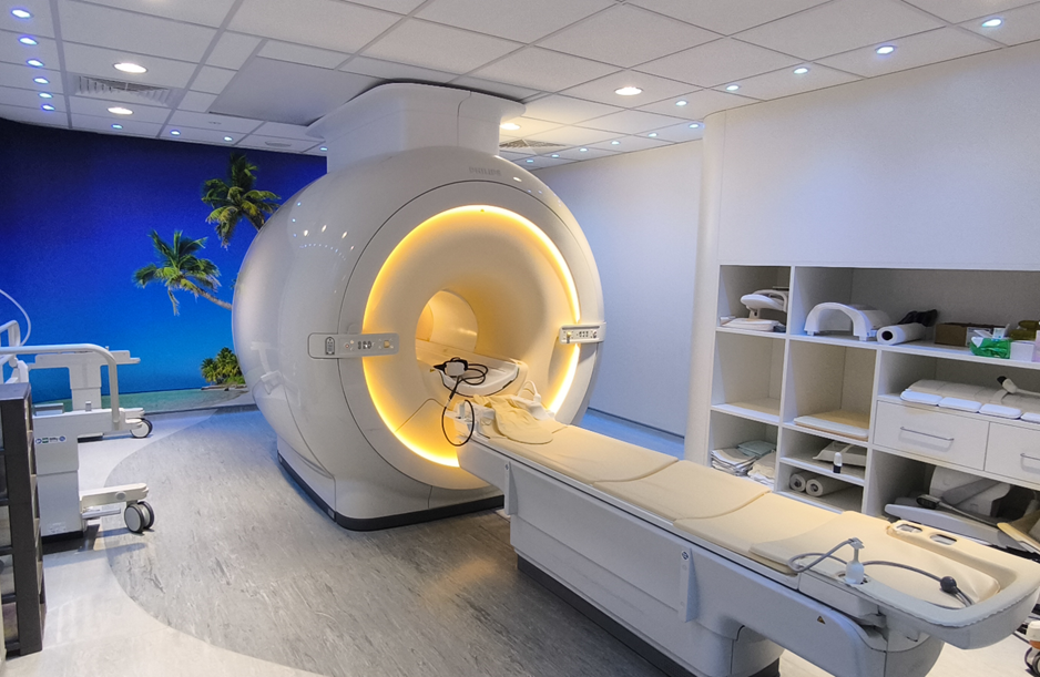
At L&D we are proud of our imaging team. We aim to provide the very best for our patients and visitors. Currently, the department has three state-of-the-art scanners located in the Main Hospital proving high resolution images to our Specialist Dr Radiologist and Cardiologist read and provide the best report.

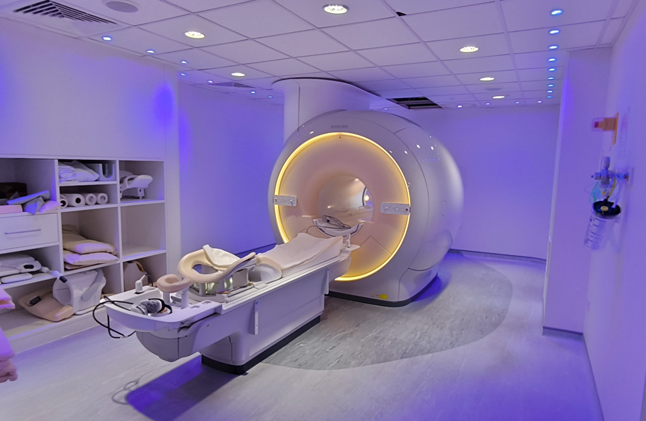
MRI Machine in a room with a beach on one wallMRI Machine in a room with purple ambient lighting

Philips Ingenia

Our facility uses the latest Philips Ingenia MRI machine which provides high resolution images with minimal noise, improving the overall accuracy of the scan.

In bore Experience

We understand that undergoing an MRI scan can be a daunting experience. That is why, we provide a calm and comfortable environment, with experienced Radiographers who will guide you through the entire process. In one of our scanners we can provide a TV experience where depending on each type scan you will have, you can have access to see some animation themes in the screen though a mirror.

Please ensure that you arrive at least 15 minutes before your appointment at MRI department, so that no inconvenience is caused to yourself or other patients.

  • Bring a Companion: If you feel anxious or claustrophobic, consider bringing a trusted companion to support you throughout the procedure. Also, request your GP for some type of tranquilizer.
  • Dress Comfortably: Wear comfortable clothing without metal zippers, snaps or buttons, as metal can interfere with the MRI machine. We will provide you with a gown to wear during the scan.
  • Remove Accessories: Remove all jewellery, watches, and hairpins before the scan, if possible come prepared, this will help with the workflow. We provide a locker for your essential belongings, but we cannot take any responsibility for it in the event of loss.
  • Avoid Food and Drink: Avoid large meals before your scan, and follow the instructions we send in the letter, different type of scan have different preparation.

The machine takes a few minutes to capture each image, so it is important to lie as still as possible throughout the procedure. You will be provided with headphones or hear plugs to protect your hearing and essential equipment during your scan, music will be provided through your headphones only to listen to during the scan, to help you relax and pass the time. Some scans will be asked some breathing instruction, the Radiographer will speak with you before scan starts and do some practice.

Very occasionally people can become too warm during an MRI scan, this is the main reason our MRI rooms are maintained in low temperature and the MRI scanners have some active ventilation.

For some MRI scans it will be necessary for the Radiographer to give you an injection of contrast media into a vein, through a cannula (soft, thin plastic tube) during your scan. This is a clear, colourless fluid that is used to make the images clearer during an MRI scan and help with diagnosis.

The contrast is a Gadolinium-based contrast agents (also known by their brand names Gadovist® Prohance® (Gadoteridol) and Primovist® (Gadoxetic disodium), which may be used during MRI scans. If you have any questions or concerns, please speak to your doctor or nurse. Further information about the contrast agent is available in the manufacturer’s patient information leaflet – please ask us if you would like a copy.

Side effects

The contrast is not suitable for everybody. The radiographer or radiologist will decide if it is appropriate for you.

You must tell the radiographer or radiologist before your scan if you have any history of poor renal function or kidney problems, or if you have ever had an allergic reaction to a contrast agent.

The contrast does not usually cause any reaction. Some of the more common reactions include:

  • Injection site pain; Nausea; Vomiting; Itching; Rash; Headache; Paraesthesia (abnormal skin sensation, such as prickling, burning or tingling). Swelling of the face, mouth, hands, feet or throat; difficulty in breathing or swallowing; fainting; coughing, wheezing, and sneezing; eye irritation rash, hives, itchy red skin.

The reactions usually occur during the injection or within the first hours afterwards; however, some can occur up to several hours later.

If you have any concerns about side effects, please speak to the staff caring for you.

Breastfeeding Patients

A small amount of the contrast agent is excreted into the breast milk. There is no proven risk to the infant, but if you are worried you could discard breast milk for 24 hours after the injection.

Contact us

If you have any questions or concerns, or if you experience any of the symptoms listed above, please contact the MRI Department:

  • T: 01582 497438 (Monday to Friday, 9am-5pm)
  • Out of hours, please contact your GP or go to your local Emergency Department (A&E)

Results

Once the MRI scan is complete, our Dr Radiologist will interpret the images and prepare a report to send to your referral.

Follow-Up

It is important to follow-up with your healthcare provider to discuss your MRI results and determine any necessary next steps.

Routine Scans

We provide a large range of routine scans:

Brain MRI: A brain MRI is done to check for tumours, an aneurysm, bleeding in the brain, nerve injury, or other brain problems.

Scan of a brain using an MRI machine

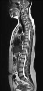
Spine MRI: A spine MRI is done to check for herniated discs, spinal cord injuries, tumours, or other spine problems.

Scan of a spine using an MRI machine

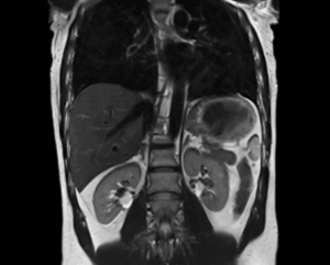
Abdominal MRI: An abdominal MRI is done to check for tumours, bleeding, infection, or other problems in the abdomen.

Scan of a abdominal cavity using an MRI machine

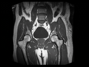
Pelvic MRI: A pelvic MRI is done to check for tumours, infection, bleeding, or other problems in the pelvis.

Scan of a pelvis using an MRI machine

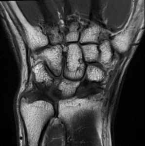
Musculoskeletal System: MSK examinations are exams to extremities of your body. Arms, hands, legs, foot.

Scan of a Musculoskeletal System using an MRI machine

Specialized Scans

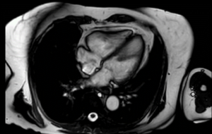
Cardiac Scan: A Cardiac scan it is done to check the morphology and function of the Heard. We have a specialized Dr Cardiologist running the list with the specialized Radiographer.

A cardiac scan using an MRI machine

Cardiac Stress Scan: We use a drug for you to you have the same sensations as you are doing some exercise and at that time you have the MRI scan done.

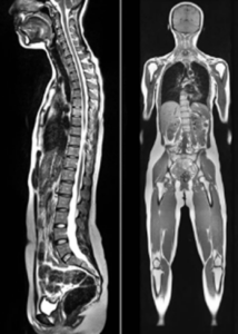
Whole body: It is a very complex scan witch takes sometimes more than one hour. For this scan all your body will be covered for MRI coils and we just ask you to be very still.

A whole body scan using an MRI machine

Defecating Proctogram: This is a very specialized scan, which needs to be referred by a specific clinic and approved by a Senior Radiologist.

If you have any medical devices in your body, you need to contact us before your appointment. The team need to analyse the device brand and model to assess scan safety.

There is a chance, even if your device is branded as MRI-safe, that you won’t be able to have your procedure with us. This can be dependent on your device type and features.

Pacemakers:

We can perform a MRI scan on a patients having a Pacemaker. Essential information need to be provided by you and your referral:

  • The Pacemaker passport with brand and model for the box and wires
  • Last Pacemaker check report
  • Chest X-Ray within three months

When provided with the information, the team will assess whether you are able to have your scan with us.

Please note: We just scan Systems MRI Safety.

If you are external referral or your Pacemaker System has been done outside of L&D please contact us.

Some devices we are not able to scan in our department:

ICD (Implantable cardioverter-defibrillator) – Conditional and non-Conditional.

Devices non-MRI conditional.

Devices requiring Physicist supervision.

We have two different locations with MRI scans, please make sure you follow your letter address indications.


MRI at Luton and Dunstable Hospital

The Hospital is situated just off junction 11 of the M1 Motorway.

The Imaging department is situated on the 1st floor of the Medical Block.

Parking: There is a carpark in opposite the main entrance in the Lewsey Road. There is a charge for parking.


MRI mobile scanner at Grove View Health Hub

Opening times: 8am – 8pm, 7 days a week.

The MRI mobile scanner at Grove View is provided by a private company allied with our MRI department. The same MRI scans are used here as in the main department. The company use the same protocols and we closely monitor the examinations quality.

The MRI is situated behind the main entrance of the building. On your arrival, please knock and show the Radiographer your letter and completed safety questionnaire.

Parking

Parking is available on site free for two hours. You will need to register your car in the main entrance of the Hub.

Facilities

The nearest toilets are in the main Hub.

Mobility issues

Unfortunately, we do not have a hoist at this location. We advise patients with mobility issues not to attend. The mobile has a staircase and lift for patients to access them.